Respiratory syncytial virus (RSV) is a common respiratory infection that can be serious for infants...
Some features of our site, including online booking, are temporarily unavailable while we work on improving your care experience! Please call to schedule.
If you experience lung pain or discomfort, your doctor may recommend pulmonary endoscopy to determine the source of your symptoms.
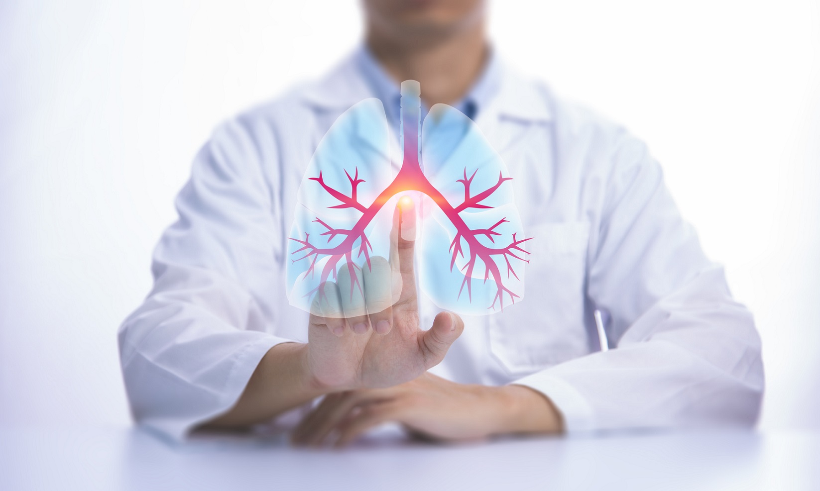
Pulmonary endoscopy, also known as bronchoscopy, is a procedure that helps doctors visualize and examine the inside of your airways and lungs. Using a bronchoscope—a flexible tube with a camera and light—your doctor can diagnose and treat various respiratory conditions directly. This minimally invasive procedure is crucial for identifying abnormalities and administering treatments within the bronchial passages and lungs.
Pulmonary endoscopy helps diagnose various respiratory issues, such as chronic cough, unexplained infections, and abnormal chest imaging results. Conditions like lung cancer, bronchiectasis, and interstitial lung disease can be effectively diagnosed and managed through this procedure. Beyond diagnosis, pulmonary endoscopy can also facilitate therapeutic interventions like clearing airway obstructions, removing foreign bodies, and delivering medications directly to the lungs.

Your physician may perform an endobronchial ultrasound (EBUS) bronchoscopy to better examine your bronchi—the airways leading to your lungs. This diagnostic procedure is conducted using a bronchoscope equipped with an ultrasound probe to create images of your lungs and surrounding lymph nodes. EBUS bronchoscopy allows pulmonologists to accurately assess areas of concern from previous scans or X-rays that require a closer look.

This procedure uses a special type of bronchoscope to reach areas of the lungs that traditional bronchoscopes cannot access. Using electromagnetic technology and real-time computerized tomography (CT), doctors can generate a three-dimensional map of the lungs. With this guide in hand, they can access hard-to-reach regions of the lung that require removal or targeted radiation. Robotic Bronchoscopy is also available, providing your pulmonary specialist with state-of-the-art tools to diagnose various conditions.
Bronchoscopy is conducted with a device that allows doctors to see inside your body. An endoscope is a long, thin tube with a camera and a light attached to one end. It also includes an open channel through which medical tools can pass to collect tissue samples for biopsies.
A bronchoscope can be inserted through the nose or mouth. A local or general anesthetic will be administered to encourage relaxation and reduce discomfort during the procedure. In addition to taking samples, a bronchoscopy can also be used to remove tumors in the lung via cutting tools, electric currents, lasers, or light-sensitive chemicals.
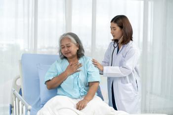
After a pulmonary endoscopy, patients are monitored until the sedative wears off. Common symptoms like a sore throat, hoarseness, and mild cough usually resolve within a few days. Normal activities can usually be resumed within 24 hours. If a biopsy was done, there may be a slight risk of complications like bleeding or infection. Contact your doctor if you experience severe pain, fever, or persistent bleeding.

Respiratory syncytial virus (RSV) is a common respiratory infection that can be serious for infants...
Zephyr valves are tiny, one-way devices that help people with severe COPD or emphysema breathe...

Pneumonia is a lung infection that can range from mild to life-threatening. Learn the common causes...

Learn more about our online scheduling and schedule an appointment with your primary care provider today.

We offer a wide variety of services at our many locations throughout New Jersey, including award-winning obstetrics and gynecology, cancer care and orthopedics.

World class care is in your backyard. Learn more about our local and nationally renowned physicians.