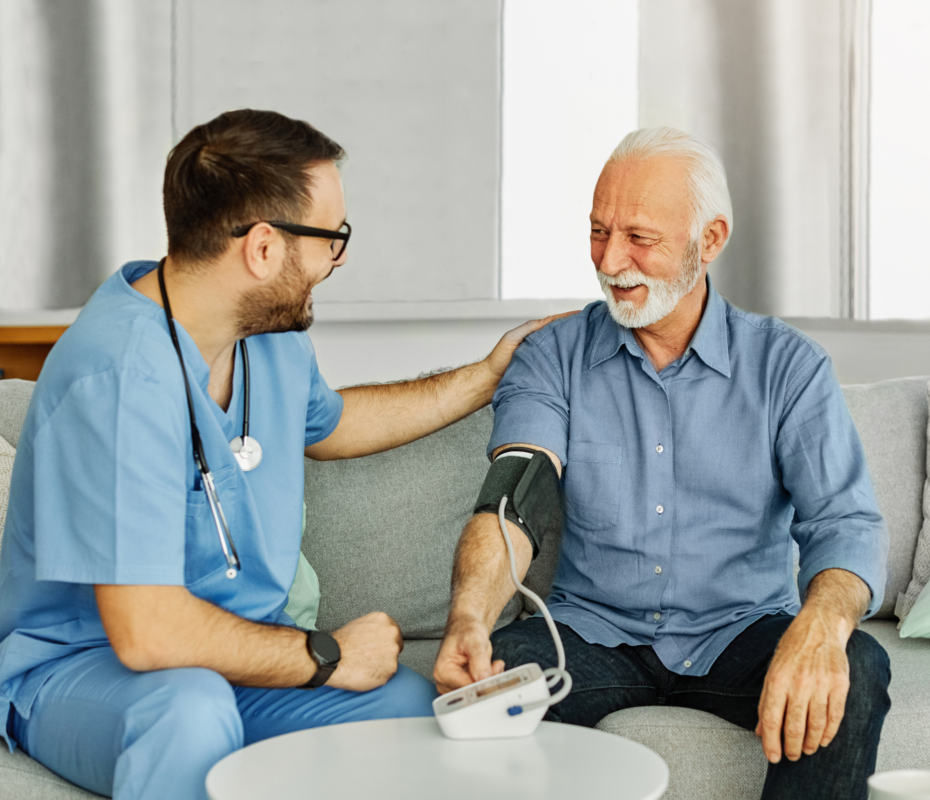
Paul Maurice turned to Inspira Rehab Services for pain relief and mobility after an ankle injury. He...
Some features of our site, including online booking, are temporarily unavailable while we work on improving your care experience! Please call to schedule.
When blood vessels become narrowed or blocked, it can lead to serious health problems like carotid artery disease that may lead to a stroke. An angiogram is a diagnostic imaging test that helps doctors see how blood flows through your vessels and identify any areas of concern.
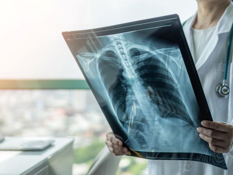
An angiogram, also known as an arteriogram, is a medical imaging procedure used to visualize the inside of blood vessels and organs, particularly arteries and veins. By injecting a special contrast dye into the bloodstream, an angiogram allows doctors to see detailed X-ray images of how blood flows through the vessels. This procedure helps identify blockages, narrowing or other abnormalities that may be causing symptoms or increasing the risk of serious conditions.
An angiogram helps diagnose a variety of vascular conditions, including coronary artery disease, peripheral artery disease (PAD), aneurysms, blood clots, renal artery stenosis, mesenteric ischemia and blood vessel malformations. By pinpointing areas of concern, it helps guide treatment decisions, including whether medications, lifestyle changes or surgical interventions can help improve blood flow and prevent complications.
An angiogram is a specialized imaging test that uses X-ray technology and a contrast agent (or in some cases CO₂ gas) to create detailed pictures of your blood vessels. By highlighting blood flow in real time, it helps doctors identify narrowing, blockages, aneurysms or other vascular abnormalities. In some cases, treatment such as angioplasty or stent placement can be performed immediately after the angiogram to restore healthy circulation.
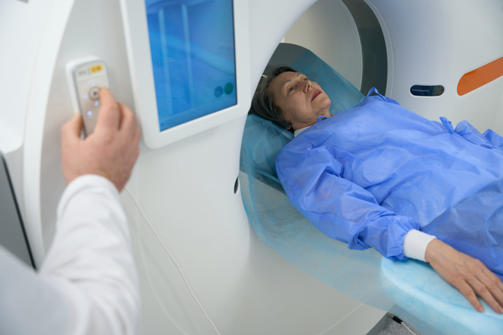
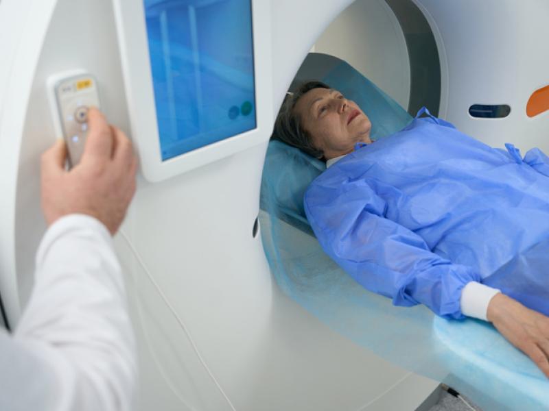
There are several types of angiograms: a traditional catheter-based angiogram, or less invasive computed tomography (CT) and magnetic resonance (MR) angiograms. The type of angiogram you have depends on your needs and condition.
A coronary angiogram examines the arteries that supply blood to the heart. It helps detect blockages or narrowing that could lead to chest pain, heart attack or other heart conditions.

A cerebral angiogram provides detailed images of the blood vessels in the brain. It can identify aneurysms, vascular malformations or signs of stroke.
A pulmonary angiogram looks at the blood vessels in the lungs. It can diagnose a pulmonary embolism (PE) , which is a potentially life-threatening blood clot.
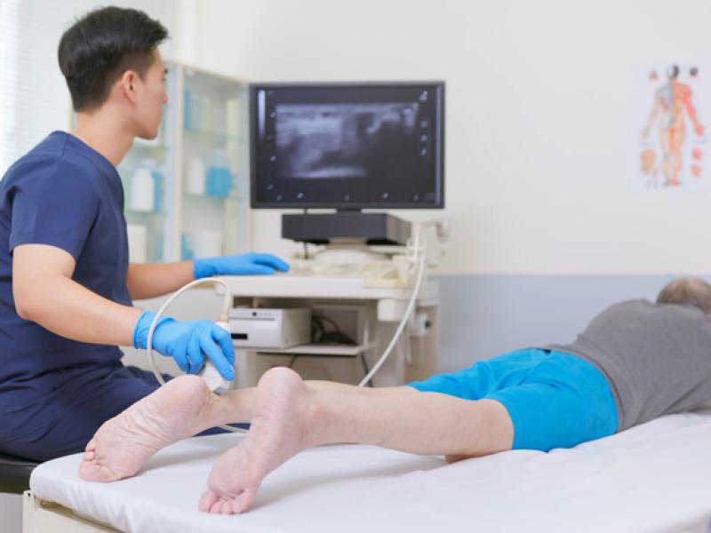
A peripheral angiogram evaluates blood flow in the arteries of the arms, legs, hands or feet. It helps diagnose PAD and may include imaging of specific arteries, such as the femoral artery in the thigh.
A renal angiogram examines the arteries that supply blood to the kidneys. It can help diagnose conditions such as renal artery stenosis, which may contribute to high blood pressure or reduced kidney function.
A mesenteric angiogram images the arteries that supply the intestines. It can identify narrowed or blocked vessels that may be causing abdominal pain or digestive problems.
Before your angiogram, you'll receive specific instructions about fasting, medications and allergy precautions—especially related to contrast dye or iodine. Pre-procedure tests like blood work or imaging may also be required. On the day of the procedure, you'll change into a gown and an IV will be placed to administer fluids and sedation.
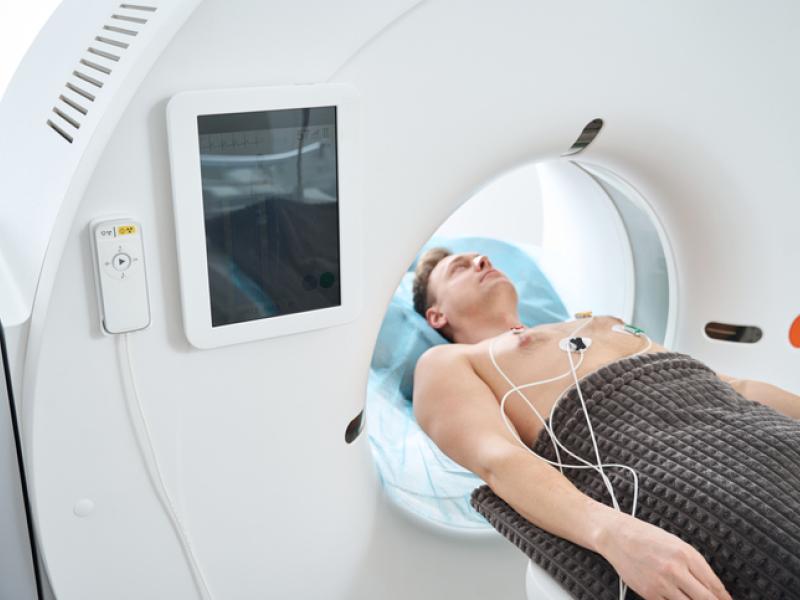
During the angiogram, you'll remain awake but relaxed with a mild sedative. The doctor will numb the access site—typically your groin, wrist or arm—before inserting a thin catheter into a blood vessel. Using X-ray imaging and contrast dye, they’ll examine blood flow and detect any blockages or abnormalities. You may feel slight pressure, but the procedure is generally painless and lasts 30 minutes to a few hours.
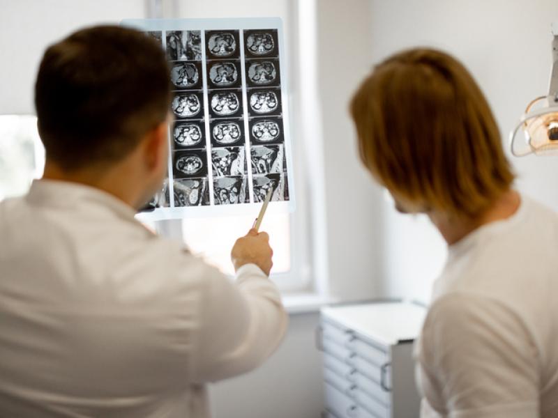
Afterward, the catheter is removed and pressure applied to the site to prevent bleeding. You'll be monitored as the sedation wears off. Most patients return home the same day, though some may stay overnight. Recovery includes resting, avoiding heavy lifting, and watching for signs of infection or bleeding. Your doctor will follow up with results and discuss any necessary treatments.

Paul Maurice turned to Inspira Rehab Services for pain relief and mobility after an ankle injury. He...

Blood clots can happen to anyone, even elite athletes. Here’s what to know about how blood clots...

Varicose veins are twisted, enlarged veins that can cause discomfort, swelling and circulatory...

Learn more about our online scheduling and schedule an appointment with your primary care provider today.

We offer a wide variety of services at our many locations throughout New Jersey, including award-winning obstetrics and gynecology, cancer care and orthopedics.

World class care is in your backyard. Learn more about our local and nationally renowned physicians.