Paul Maurice turned to Inspira Rehab Services for pain relief and mobility after an ankle injury. He...
Some features of our site, including online booking, are temporarily unavailable while we work on improving your care experience! Please call to schedule.

An aortic aneurysm develops when your main artery weakens and bulges outward under pressure. Often symptomless at first, it can become life‑threatening if the bulge grows and ruptures, making regular monitoring and timely care essential.
An aortic aneurysm is a localized dilation of the body’s main artery caused by a weakness in the vessel wall. Over time, factors like high blood pressure, atherosclerosis, smoking and certain genetic disorders contribute to gradual wall degeneration. As the aneurysm enlarges, the risk of aneurysm rupture increases, sometimes leading to catastrophic internal bleeding and shock. In some cases, an arterial wall infection can result in a mycotic aneurysm or pseudoaneurysm, which carry their own unique risks.
There are several types of aortic aneurysms based on location and shape:
A dilation of the abdominal segment of the aorta, often detected by ultrasound screening to prevent rupture
A bulge in the chest portion of the aorta that can progress to an aortic dissection if the inner wall tears
An aneurysm spanning both the thoracic and abdominal aorta, requiring careful surgical planning
A localized, pouch‑shaped outpouching of the aortic wall, increasing rupture risk at the narrow neck
A uniform, spindle‑shaped dilation involving the entire circumference of the aorta
A tear in the inner layer of the aortic wall allows blood to split its layers, causing severe pain
A contained rupture where blood collects between vessel layers or surrounding tissue rather than within all three aortic wall layers
Other types of aneurysms include peripheral aneurysms (popliteal or femoral) and mesenteric aneurysms, which occur in arteries outside the aorta.
Many aortic aneurysms remain symptomless until they grow large or rupture, but possible warning signs include:
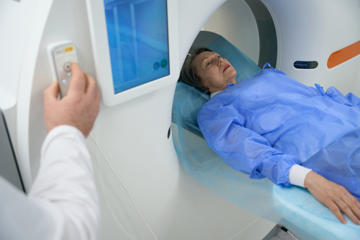
The doctor may begin by visualizing vessel size and wall integrity. If detected, computed tomography angiography (CTA) provides high‑resolution, cross‑sectional images that confirm diameter, shape and the involvement of branch vessels. Magnetic resonance angiography (MRA) offers a radiation‑free alternative, which is particularly useful in patients with contrast allergies or kidney issues. Invasive digital subtraction angiography can map complex anatomy before endovascular repair.
Thoracic endovascular aneurysm repair (TEVAR) and EVAR are treatments that use a catheter in the groin to deliver a stent graft to reinforce the weakened section of the aorta. EVAR is used for abdominal aneurysms and TEVAR for thoracic aneurysms, offering a less invasive alternative to open surgery with significantly shorter recovery times.
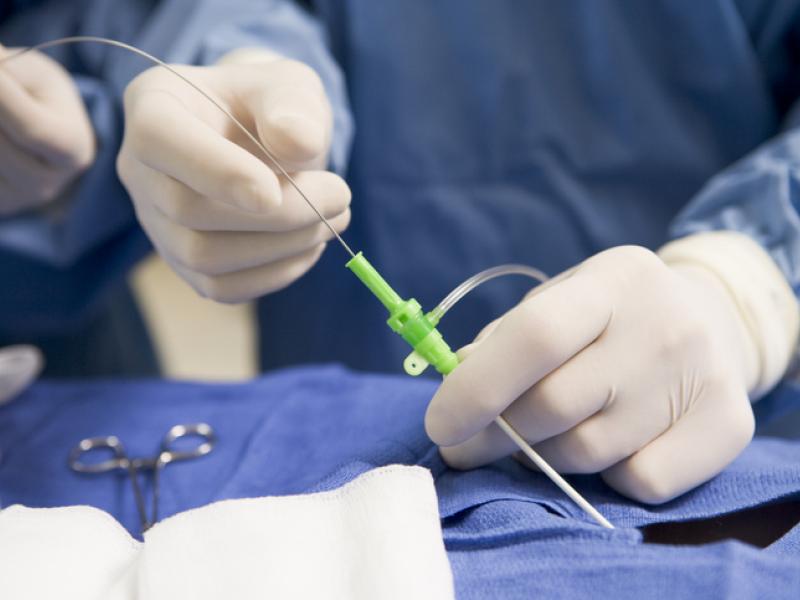
For aneurysms involving branch arteries or unusual vessel shapes, fenestrated stent grafts, with built‑in openings that align to side branches, and branched stent grafts, which incorporate side‑arms to vital vessels, preserve critical blood flow without open surgery. In the most complex cases, hybrid procedures can achieve a customized repair with minimal invasiveness.

Small aneurysms may not require surgery and are monitored with regular imaging. Blood pressure, cholesterol and lifestyle factors are monitored and managed to slow aneurysm growth.

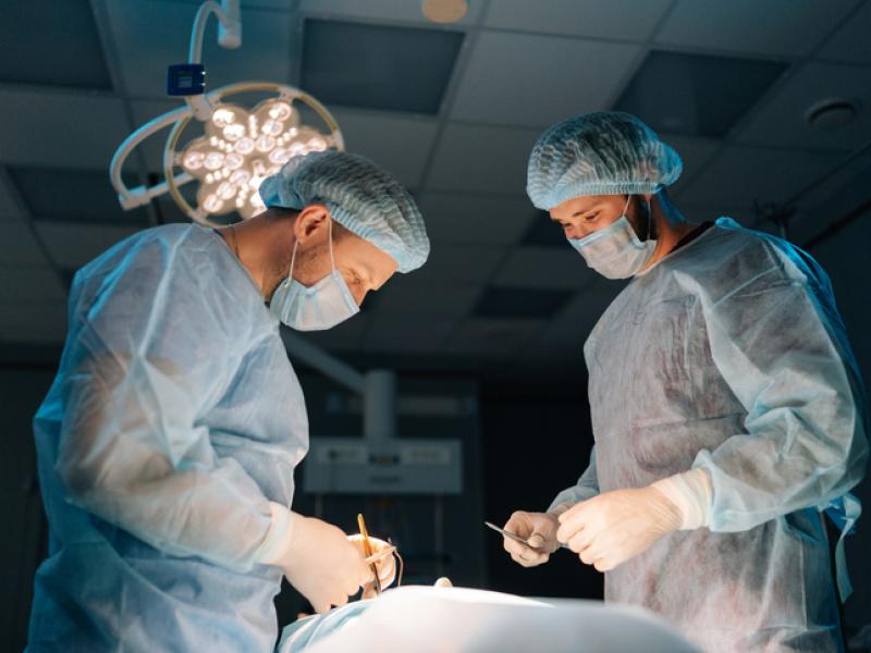
The surgeon makes an incision in the abdomen or chest to remove the aneurysm and replaces it with a synthetic graft. This approach provides a durable solution for large or complex aneurysms that may not be suitable for less invasive, endovascular techniques.
Inspira offers a comprehensive, team‑based approach to aortic aneurysm care, bringing together vascular surgeons, interventional radiologists, cardiologists, specialized nursing staff and more to deliver personalized treatment plans. From initial screening and diagnostic imaging through follow‑up surveillance, our coordinated care ensures you receive the right intervention. We use advanced imaging technology and evidence‑based protocols to monitor aneurysm size and growth, adjusting treatment recommendations to minimize risk and optimize outcomes.
With convenient access to care at multiple locations across South Jersey, we strive to make your experience as seamless and reassuring as possible, because managing aortic aneurysms is not just about treating the vessel, but supporting the whole person.
Paul Maurice turned to Inspira Rehab Services for pain relief and mobility after an ankle injury. He...

Blood clots can happen to anyone, even elite athletes. Here’s what to know about how blood clots...

Varicose veins are twisted, enlarged veins that can cause discomfort, swelling and circulatory...

Learn more about our online scheduling and schedule an appointment with your primary care provider today.

We offer a wide variety of services at our many locations throughout New Jersey, including award-winning obstetrics and gynecology, cancer care and orthopedics.

World class care is in your backyard. Learn more about our local and nationally renowned physicians.