Paul Maurice turned to Inspira Rehab Services for pain relief and mobility after an ankle injury. He...
Some features of our site, including online booking, are temporarily unavailable while we work on improving your care experience! Please call to schedule.

An abdominal aortic aneurysm (AAA) happens when a section of the abdominal aorta becomes weakened and bulges outward. While often symptomless at first, abdominal aortic aneurysms can become life-threatening if they grow too large or rupture without warning.
An abdominal aortic aneurysm (AAA) is when the abdominal portion of the aorta, the main blood vessel that carries blood from the heart to the lower body, balloons. The aorta normally has thick, elastic walls that withstand high pressure, but certain factors can weaken its structure over time. Early mild widening of the vessel, called an ectatic aorta, can progress to an aneurysm when the diameter exceeds 1.5 times its regular size.
AAAs can be classified by their location in relation to the renal arteries, blood vessels that supply the kidneys. Each type has different implications for monitoring and treatment based on how close the aneurysm is to vital branches of the aorta.
Several key factors raise the risk of developing an abdominal aortic aneurysm. Age over 65 is the strongest risk factor. People who smoke are especially at risk, as smoking damages aortic walls. High blood pressure and a family history of aneurysm also increase risk. Early detection and understanding personal risk can help prevent serious complications.
AAAs often develop slowly and silently, with no symptoms until the aneurysm becomes large or ruptures. When symptoms do occur, they may include:
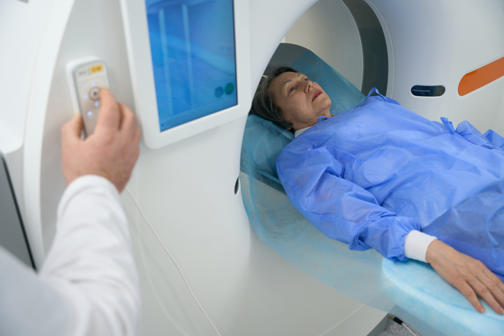
During AAA screening, your doctor will check your abdomen for a pulsating mass. If detected, an ultrasound can confirm the presence and detect aneurysm from 3 centimeters and bigger. For more detailed images, CT angiography or MRI may be used to assess the aneurysm’s size, shape and relationship to nearby renal and iliac vessels, helping guide your treatment plan.
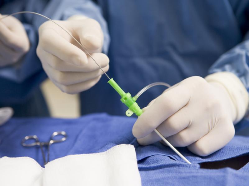
A minimally invasive procedure in which a stent graft is delivered through the femoral arteries to reinforce the weakened aorta from within, shortening recovery time and lowering perioperative risk.

Traditional AAA repair surgery involves an incision in the abdomen, direct removal of the aneurysmal segment and replacement with a synthetic graft. This procedure offers durable, long-term results for large or complex AAAs.

Moderate size AAAs (typically less than 5.5 centimeters in diameter) are monitored with periodic AAA ultrasound exams every six to 12 months to track growth and decide when intervention is needed.

Managing high blood pressure with medications, lowering cholesterol with statins and quitting smoking can slow aneurysm growth and boost overall cardiovascular health.
Our multidisciplinary team of vascular surgeons, interventional radiologists, primary care doctors and cardiologists guides you through AAA care from screening to recovery. We offer the latest minimally invasive procedures and personalized treatment plans. Our experts focus on safety, proven results and ongoing support, helping you understand options and maintain your health at every stage.

Paul Maurice turned to Inspira Rehab Services for pain relief and mobility after an ankle injury. He...

Blood clots can happen to anyone, even elite athletes. Here’s what to know about how blood clots...

Varicose veins are twisted, enlarged veins that can cause discomfort, swelling and circulatory...

Learn more about our online scheduling and schedule an appointment with your primary care provider today.

We offer a wide variety of services at our many locations throughout New Jersey, including award-winning obstetrics and gynecology, cancer care and orthopedics.

World class care is in your backyard. Learn more about our local and nationally renowned physicians.