Paul Maurice turned to Inspira Rehab Services for pain relief and mobility after an ankle injury. He...
Some features of our site, including online booking, are temporarily unavailable while we work on improving your care experience! Please call to schedule.

Aortic dissection is a rare but life-threatening condition that happens when the inner layer of the aorta tears, allowing blood to flow between the layers of the vessel wall. This condition can lead to severe complications if not treated quickly, making early recognition and emergency care essential.
Aortic dissection is a serious condition that happens when a tear develops in the inner layer of the aorta, the large artery that carries blood from the heart to the rest of the body. When the aorta tears, blood surges through the opening, separating (or dissecting) the inner and middle layers of the aortic wall.
In some cases, advanced atherosclerotic plaque can erode through the aorta’s inner layer, forming a penetrating aortic ulcer that may extend into the middle layer. When either an aortic tear or penetrating ulcer occurs, blood surges through the opening, separating (or dissecting) the inner and middle layers of the aortic wall. This separation creates a false channel for blood flow, which can reduce or block blood supply to vital organs and may lead to aortic rupture or stroke if not treated quickly.
Aortic dissection types are classified by the tear's location and extent:
Aortic dissection risk increases with chronic high blood pressure, connective tissue disorders like Marfan syndrome or Ehlers-Danlos, and preexisting aortic aneurysms. Other causes include atherosclerosis and chest trauma. The condition is most common in men and people assigned male at birth between ages 60 and 80, though genetic factors can lead to earlier onset.
Symptoms of aortic dissection often come on suddenly and can include:
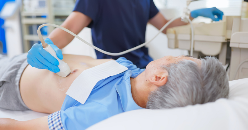
Early diagnosis of aortic dissection is lifesaving. Doctors begin with a physical exam and review of symptoms such as chest pain and unequal blood pressure between arms. The most common test is a CT angiogram, which provides detailed images of the aorta and the tear’s severity. Other options include a transesophageal echocardiogram (TEE) or MRI for patients who cannot receive contrast dye.

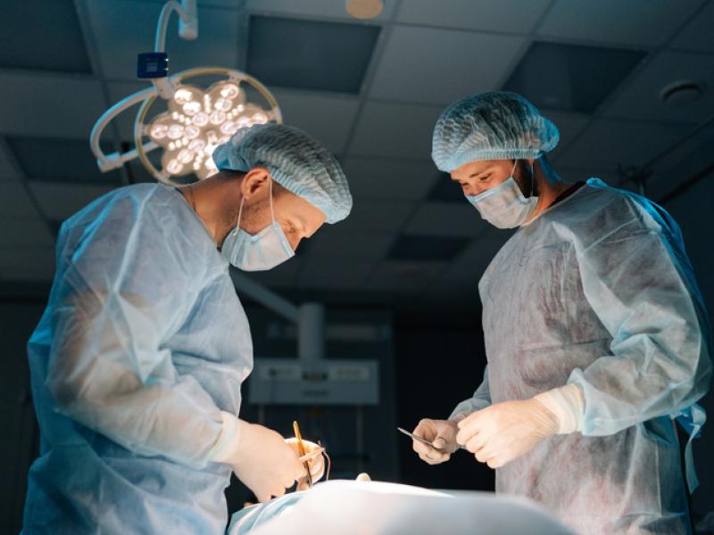
In complex dissections, a combination treatment of open surgery and endovascular techniques can tailor the repair to your body while reducing overall invasiveness.

Initial treatment focuses on controlling blood pressure to reduce stress on the aortic wall and prevent further tearing. Pain control with opioid medications can alleviate discomfort and further stabilize hemodynamics.

For dissections involving the ascending aorta (Stanford type A) or complicated type B cases, open surgery can remove the damaged portion and replace it with a synthetic graft.
Endovascular stent grafting is a minimally invasive procedure that seals the tear and restores normal blood flow. Thoracic endovascular aortic repair (TEVAR) is performed in the chest, and endovascular aneurysm repair (EVAR) is performed below the diaphragm in the abdominal aorta.

Lifelong imaging follow-up (CT or MRI) is required after any intervention to monitor for graft integrity and detect late complications. Continued blood pressure management and lifestyle modifications can help prevent progression or recurrence.
At Inspira, our vascular specialists work closely with cardiothoracic surgeons, interventional radiologists and critical care teams to deliver fast, coordinated care for aortic dissection. From emergency imaging and medical stabilization to minimally invasive endovascular repair or open surgery, every step is managed with precision and urgency.
After treatment, we focus on long-term recovery through routine imaging, blood pressure control and lifestyle support. Our goal is to help you heal, stay healthy and regain confidence with expert care and ongoing guidance.
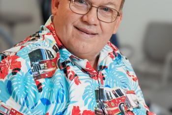
Paul Maurice turned to Inspira Rehab Services for pain relief and mobility after an ankle injury. He...
Blood clots can happen to anyone, even elite athletes. Here’s what to know about how blood clots...

Varicose veins are twisted, enlarged veins that can cause discomfort, swelling and circulatory...

Learn more about our online scheduling and schedule an appointment with your primary care provider today.

We offer a wide variety of services at our many locations throughout New Jersey, including award-winning obstetrics and gynecology, cancer care and orthopedics.

World class care is in your backyard. Learn more about our local and nationally renowned physicians.