Paul Maurice turned to Inspira Rehab Services for pain relief and mobility after an ankle injury. He...
Some features of our site, including online booking, are temporarily unavailable while we work on improving your care experience! Please call to schedule.

A thoracic aortic aneurysm is a bulge or weakening in the wall of the aorta (the body's largest artery) as it passes through the chest. While it often develops slowly and without symptoms, a thoracic aortic aneurysm can become life-threatening if it ruptures or leads to a tear in the aorta.
A thoracic aortic aneurysm (TAA) is an abnormal bulge or dilation of the aorta (body's largest artery) that runs through the chest. Over time, the wall of the aorta weakens, often due to genetic factors, high blood pressure or damage to the arterial lining, allowing it to stretch under the force of blood flow. Most TAAs develop slowly and may not cause noticeable symptoms until they reach a size or shape that threatens the integrity of the vessel wall.
There are several types of TAAs, classified by their location and shape:
Long-term high blood pressure weakens the aortic wall, while genetic conditions like Marfan syndrome or a bicuspid aortic valve reduce structural strength. Atherosclerosis, smoking and family history further damage the vessel lining and increase susceptibility to aortic disease.
TAAs often develop without noticeable symptoms. When they do occur, they may include:
Diagnosing a TAA involves imaging studies such as transthoracic or transesophageal echocardiography. CT angiography and MRI can provide high-resolution cross-sectional views of the chest aorta to determine the aneurysm’s size. Ultrasounds can also track aneurysm growth over time. Once detected, having an imaging test at regular intervals can confirm progression and guide decisions about surgical or endovascular repair.
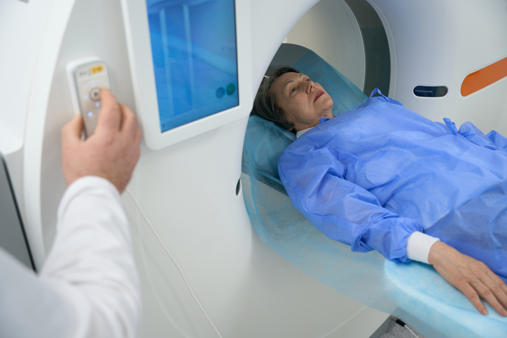

Your doctor may recommend regular imaging to monitor the aneurysm’s size and growth for small, asymptomatic TAAs. Managing blood pressure, avoiding tobacco and making heart-healthy lifestyle changes can help slow progression.

Medications like beta blockers or angiotensin receptor blockers (ARBs) can lower blood pressure and reduce stress on the aortic wall. These treatments can effectively slow aneurysm growth, especially in people with underlying conditions like Marfan syndrome.
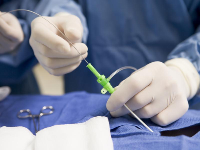
TEVAR is a minimally invasive procedure that involves placing a stent graft through a catheter in the groin to reinforce the weakened section of the aorta. TEVAR may be recommended for patients with specific aneurysm sizes, shapes or health risks that make open surgery too risky.

If a TAA ruptures or is at high risk of imminent rupture, emergency surgery, either open repair or TEVAR, is necessary to save the patient’s life. Outcomes depend on how quickly the condition is identified and treated.

Open surgery may be an option in cases where TEVAR isn’t possible or the aneurysm is large or symptomatic. A surgeon removes the damaged portion of the aorta and replaces it with a synthetic graft. This method can be more durable in certain situations.
Inspira brings together a dedicated team of vascular and cardiothoracic surgeons, interventional radiologists, specialized nursing staff and more who collaborate on every case. We tailor treatments based on patient preferences, aneurysm size and location, minimizing recovery time and risk. From initial diagnosis through post-procedure monitoring, our multidisciplinary team ensures each patient’s care plan reflects the latest advances in aortic disease management.
Paul Maurice turned to Inspira Rehab Services for pain relief and mobility after an ankle injury. He...

Blood clots can happen to anyone, even elite athletes. Here’s what to know about how blood clots...

Varicose veins are twisted, enlarged veins that can cause discomfort, swelling and circulatory...

Learn more about our online scheduling and schedule an appointment with your primary care provider today.

We offer a wide variety of services at our many locations throughout New Jersey, including award-winning obstetrics and gynecology, cancer care and orthopedics.

World class care is in your backyard. Learn more about our local and nationally renowned physicians.